World supplier of antibodies, antigens and kits since 2007

Other Products
CEA (Center) Rabbit pAb(AP Conjugated)
Price on request
Quantity
Ship worldwide. Contact order@enogene.com for ordering and bulk pricing or contact your local distributor.
Product Information
CategoryAntigen antibodies
ClonalityPolyclonal
ConjugationAPRecommended Apps:WB, IHC-PReactive species:HumanStore the buffer:Purified polyclonal antibody supplied in PBS with 0.09% (W/V) New type preservative N. This antibody is purified through a protein A column, followed by peptide affinity purification.
Human Gene IdNP_004354.2
SpecificityThis CEA antibody is generated from rabbits immunized with a KLH conjugated synthetic peptide between 400-429 amino acids from the Central region of human CEA.
HostRabbit
IsotypeIgG
FunctionCell surface glycoprotein that plays a role in cell adhesion, intracellular signaling and tumor progression (PubMed:2803308, PubMed:10910050, PubMed:10864933). Mediates homophilic and heterophilic cell adhesion with other carcinoembryonic antigen-related cell adhesion molecules, such as CEACAM6 (PubMed:2803308). Plays a role as an oncogene by promoting tumor progression; induces resistance to anoikis of colorectal carcinoma cells (PubMed:10910050).
DilutionWB 1:500-2000. IHC-P 1:50-200Predict molecular weight:77kDaConditions of transport and storage:Maintain refrigerated at 2-8°C for up to 2 weeks. For long term storage store at -20°C in small aliquots to prevent freeze-thaw cycles.
BackgroundCEA is a cell surface glycoprotein that plays a role in cell adhesion and in intracellular signaling. It is a receptor for E.coli Dr adhesins.Organizational expression:Expressed in columnar epithelial and goblet cells of the colon (at protein level) (PubMed:10436421). Found in adenocarcinomas of endodermally derived digestive system epithelium and fetal colon.
Cell localization:Cell membrane; Lipid-anchor, GPI-anchor. Apical cell membrane. Cell surface Note=Localized to the apical glycocalyx surface
Cell localization:Cell membrane; Lipid-anchor, GPI-anchor. Apical cell membrane. Cell surface Note=Localized to the apical glycocalyx surface
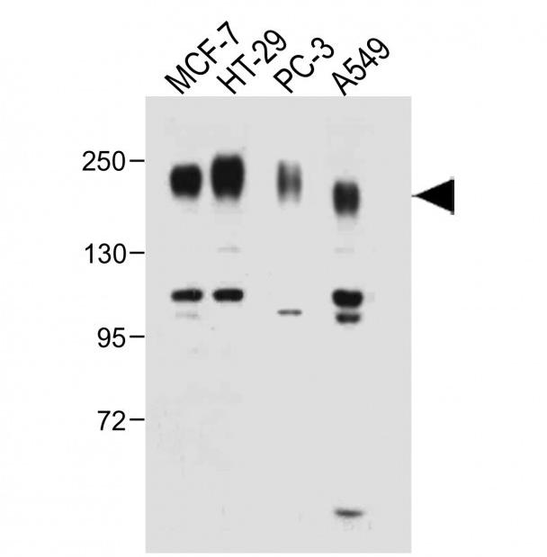
: Anti-CEA Antibody (Center) at 1:1000 dilution Lane 1: MCF-7 whole cell lysate whole cell lysate Lane 3: PC-3 whole cell lysate Lane 4: A549 whole cell lysate Lysates/proteins per lane. Secondary Goat Anti-Rabbit IgG, (H+L), Peroxidase conjugated at 1/10000 band size : 77 kD. Blocking/Dilution buffer: 5% NFDM/TBST.


Categories by research area
Categories by product type
- Antibodies
- Primary Antibodies
- Tag antibodies
- Internal control antibodies
- Pathological (Immunohistochemistry, IHC) antibodies
- Cell Signal Transduction Research Antibodies
- Secondary Antibody
- Antibody Sampler Kits
- ELISA Kits
- Recombinant Proteins
- Fluorescence Reagents
- Signal Inhibitors and Activators
- Transfection Reagent
- Cell biology kits
- Overexpression Cell Lines
- Cell Lysates
- Purification Resins
- 2019-nCoV & Monkeypox