World supplier of antibodies, antigens and kits since 2007

Cell Signal Transduction Research Antibodies
CD38 Polyclonal Antibody
Polyclonal, Rabbit
$225/ 100ul
QuantityTotal: $225
Ship worldwide. Contact order@enogene.com for ordering and bulk pricing or contact your local distributor.
Product Information
Molecular Weight34328Da
Protein NameADP-ribosyl cyclase 1
Human Gene Id952
Human Swiss Prot NoP28907
Mouse Swiss Prot NoP56528
SpecificityCD38 Polyclonal Antibody detects endogenous levels of CD38 protein.
FormulationLiquid in PBS containing 50% glycerol, 0.5% BSA and 0.02% sodium azide.
Concentration1 mg/ml
Storage Stability-20°C/1 year
Other NameCD38; ADP-ribosyl cyclase 1; Cyclic ADP-ribose hydrolase 1; cADPr hydrolase 1; T10; CD38
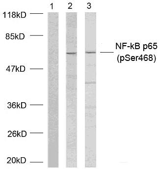
Observed Band(KD)35
Functioncatalytic activity:NAD(+) + H(2)O = ADP-ribose + nicotinamide.,developmental stage:Preferentially expressed at both early and late stages of the B and T-cell maturation. It is also detected on erythroid and myeloid progenitors in bone marrow, where the level of surface expression was shown to decrease during differentiation of blast-forming unit E to colony-forming unit E.,enzyme regulation:ATP inhibits the hydrolyzing activity.,function:Synthesizes cyclic ADP-ribose, a second messenger for glucose-induced insulin secretion. Also has cADPr hydrolase activity. Also moonlights as a receptor in cells of the immune system.,online information:CD38 entry,similarity:Belongs to the ADP-ribosyl cyclase family.,tissue specificity:Expressed at high levels in pancreas, liver, kidney, brain, testis, ovary, placenta, malignant lymphoma and neuroblastoma.,
Subcellular LocationMembrane; Single-pass type II membrane protein.
ExpressionExpressed at high levels in pancreas, liver, kidney, brain, testis, ovary, placenta, malignant lymphoma and neuroblastoma.
PurificationThe antibody was affinity-purified from rabbit antiserum by affinity-chromatography using epitope-specific immunogen.
ImmunogenThe antiserum was produced against synthesized peptide derived from the Internal region of human CD38. AA range:211-260
Gene NameCD38
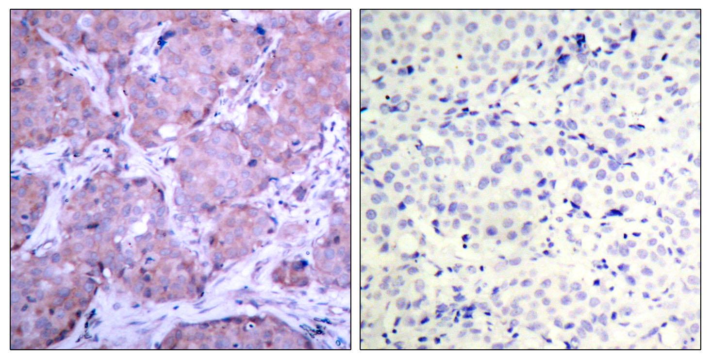
DilutionWestern Blot: 1/500 - 1/2000. IHC-p: 1/100-1/300. ELISA: 1/20000. Not yet tested in other applications.
BackgroundCD38 molecule(CD38) Homo sapiens The protein encoded by this gene is a non-lineage-restricted, type II transmembrane glycoprotein that synthesizes and hydrolyzes cyclic adenosine 5'-diphosphate-ribose, an intracellular calcium ion mobilizing messenger. The release of soluble protein and the ability of membrane-bound protein to become internalized indicate both extracellular and intracellular functions for the protein. This protein has an N-terminal cytoplasmic tail, a single membrane-spanning domain, and a C-terminal extracellular region with four N-glycosylation sites. Crystal structure analysis demonstrates that the functional molecule is a dimer, with the central portion containing the catalytic site. It is used as a prognostic marker for patients with chronic lymphocytic leukemia. Alternative splicing results in multiple transcript variants. [provided by RefSeq, Sep 2015],


Conjugated Antibody
This product is also available conjugated. Format: E20-75392-Conjugate. Price: $485 / Size: 100μl.
E20-75392-FITC$485 / 100μlE20-75392-Biotin$485 / 100μlE20-75392-HRP$485 / 100μlE20-75392-AP$485 / 100μlE20-75392-AF350$485 / 100μlE20-75392-AF405M$485 / 100μlE20-75392-AF488$485 / 100μlE20-75392-AF514$485 / 100μlE20-75392-AF532$485 / 100μlE20-75392-AF555$485 / 100μlE20-75392-AF568$485 / 100μlE20-75392-AF405S$485 / 100μlE20-75392-AF405L$485 / 100μlE20-75392-AF546$485 / 100μlE20-75392-AF594$485 / 100μlE20-75392-AF610$485 / 100μlE20-75392-AF635$485 / 100μlE20-75392-AF647$485 / 100μlE20-75392-AF680$485 / 100μlE20-75392-AF700$485 / 100μlE20-75392-AF750$485 / 100μlE20-75392-AF790$485 / 100μlE20-75392-APC$485 / 100μlE20-75392-PE$485 / 100μlE20-75392-Cy3$485 / 100μlE20-75392-Cy5$485 / 100μlE20-75392-Cy5.5$485 / 100μlE20-75392-Cy7$485 / 100μlE20-75392-PE-Cy5$485 / 100μl
Contact order@enogene.com to order conjugated formats. Magnetic Beads (1-30μm) conjugation is also available.


Categories by research area
Categories by product type
- Antibodies
- Tag antibodies
- Internal control antibodies
- Pathological (Immunohistochemistry, IHC) antibodies
- Cell Signal Transduction Research Antibodies
- Secondary Antibody
- Antibody Sampler Kits
- Recombinant Proteins
- Fluorescence Reagents
- Signal Inhibitors and Activators
- Transfection Reagent
- Cell biology kits